-

Agrisera Webinar II: Accurate Interpretation of Western Blot...
To help researchers overcome common challenges in Western Blot experiments, Xmjsci, in collaboration with Dr. Joanna, a technical ex...2025-10-23more
-
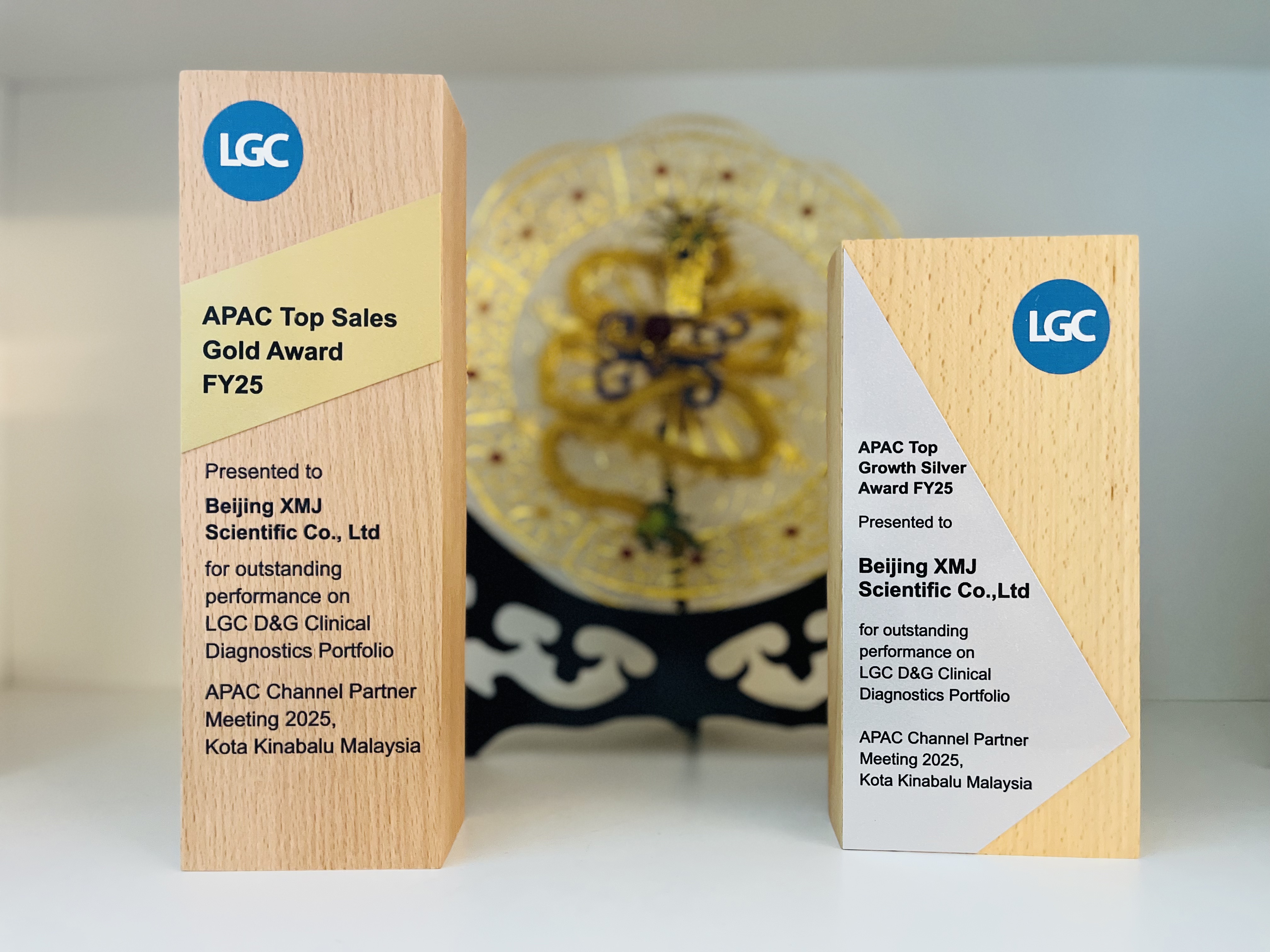
Congratulations to XMJ for winning the LGC Asia-Pacific Outs...
At the 2025 Asia-Pacific Channel Partner Conference, Beijing XMJ Scientific Co., Ltd. (hereinafter referred to as "XMJ") w...2025-10-15more
-
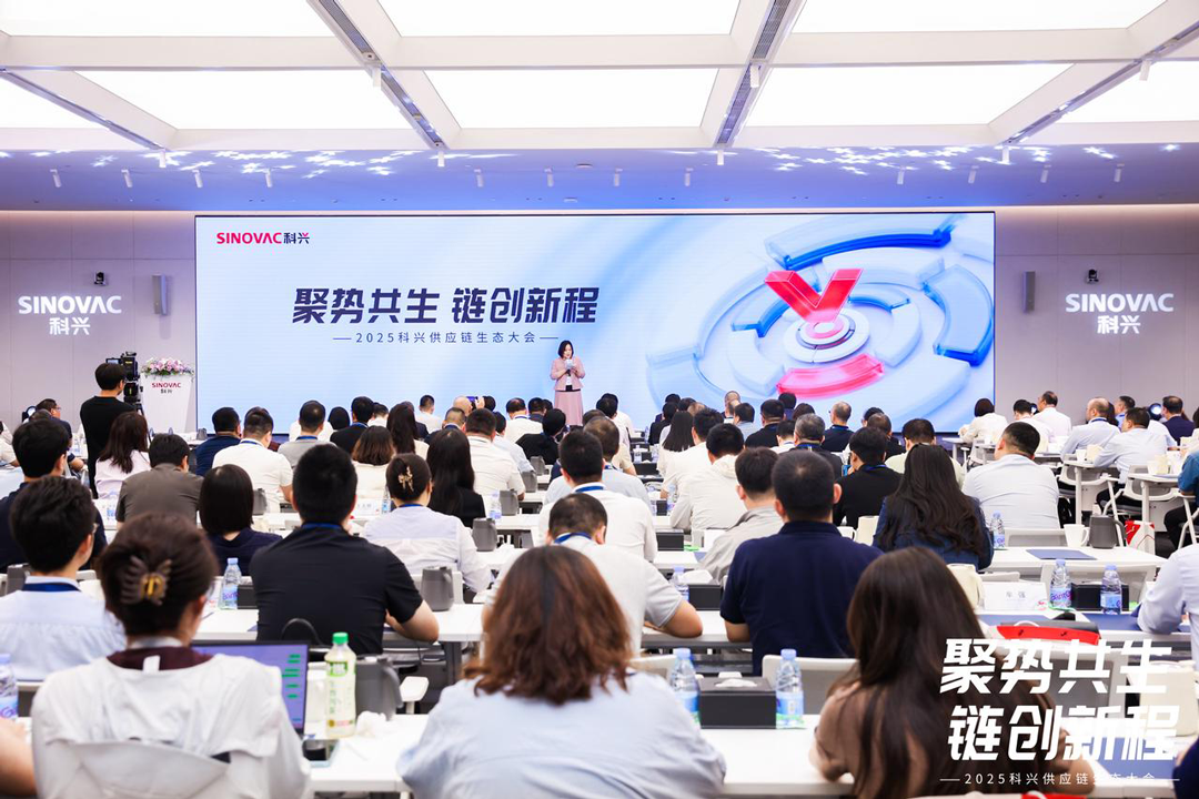
Congratulations to XMJ for being honored with Sinovac's &quo...
On August 28, the 2025 Supply Chain Ecosystem Conference, hosted by Sinovac Biotech Holdings (Group) Co., Ltd. (hereinafter referred...2025-09-01more
-

Cygnus WEBINARS | Best Practices in Host Cell Impurity Analy...
Thu, Sep 4, 2025 11:00 AM EDT (11:00 PM Asia/Shanghai)Register:Best Practices in Host Cell Impurity Analytics – Using Advanced Tec...2025-09-01more
Resouces
SeraCare?KPL Cost-Effective WB Substrate
LGC·SeraCare·KPL (hereinafter referred to as KPL), distributed by Ximeijie, is an internationally renowned manufacturer of commercially available affinity-purified secondary antibodies and substrate chromogenic systems. It has over 40 years of experience in product R&D and production, and has obtained ISO 13485 quality management system certification. Its products feature high batch-to-batch consistency and stable, reliable quality, which have won wide recognition and trust in the diagnostic reagent industry. KPL's product portfolio includes cost-effective secondary antibodies, substrate chromogenic reagents, and immunoassistive reagents, etc., which are widely used in fields such as immunological research, diagnostic reagent development, and clinical testing. KPL's cost-effective substrates and general-purpose reagents can be extensively applied to various experimental platforms, including Enzyme-Linked Immunosorbent Assay (ELISA), Western Blot (WB), Immunohistochemistry (IHC), and Enzyme-Linked Immunospot Assay (ELISpot). This article will focus on introducing the cost-effective substrate product series under the KPL brand that are suitable for Western blotting technology.Western Blot technology Western Blot technology is an immunoblotting technique used for detecting specific proteins, and it is one of the most popular methods in the field of immunoassays. Based on protein separation by molecular weight and specific antibody recognition, it involves separating proteins via electrophoresis, transferring them onto a solid-phase membrane, and then using specific antibodies for detection. In a Western Blot experiment, signal detection is a critical step, serving to visualize and quantify the target protein. In the past, colorimetric detection and low-sensitivity chemiluminescent detection drove the expansion of related research. However, with the continuous increase in requirements for detection sensitivity and advancements in imaging analysis systems, the design of chemiluminescent molecules has also achieved significant progress, enabling detection with higher sensitivity. In the Western Blot experimental system, high-quality substrates are crucial for achieving high sensitivity and low background signals. The chromogenic substrates and chemiluminescent substrates provided by KPL, with their excellent performance and reliable quality, have become core components of the Western Blot experimental system, helping researchers obtain more ideal experimental results.Chemiluminescence Chemiluminescence is the optimal choice for detecting trace proteins. Its principle lies in the luminescence of a chemiluminescent substrate when catalyzed by an enzyme (such as HRP or AP) labeled on an antibody. KPL’s LumiGLO® series consists of luminol-based HRP substrates, offering a complete range of detection solutions from regular to ultra-high sensitivity. KPL’s PhosphaGLO? series, on the other hand, are dioxetane-based AP substrates that combine high sensitivity with operational flexibility. Both series are suitable for Western Blot and ELISA applications. LumiGLO Reserve? & LumiGLO® Chemiluminescent SubstratesBoth LumiGLO Reserve? and LumiGLO® are luminol-based chemiluminescent substrates, specifically designed for high-sensitivity detection.LumiGLO Reserve?: Its sensitivity is over 20 times higher than that of LumiGLO® and other similar products, making it particularly suitable for detecting low-abundance proteins or analyzing precious samples. The luminescent signal can last for 4 to 8 hours, with the strongest signal appearing within the first hour after the reaction. The luminescence intensity is significant, allowing for easy and clear capture by chemiluminescent imagers. This product is supplied as a stable two-component solution, which can be prepared for immediate use at a 1:2 ratio. It is available in three specifications, along with matching concentrated KPL wash buffers, further enhancing experimental convenience.LumiGLO®: Also provided as a two-component solution, it can be used simply by mixing equal volumes of the two components, ensuring easy operation. In Western Blot and ELISA detection, it is more sensitive than chromogenic substrates and produces lower background. The maximum luminescence intensity is achieved within 5 minutes after reaction with HRP, and the signal can persist for 1 to 2 hours. It supports multiple stripping and re-probing of immunoblot membranes, and its linear dynamic range is superior to that of most similar products. Thus, it is widely applicable to various protein detection experiments. The detection results of both products can be permanently recorded using X-ray films or chemiluminescent imagers.Product NameItem NumberSpecificationReactive Enzyme / Catalytic EnzymeSignal TypeSensitivityLumiGLO Reserve? Chemiluminescent Substrate5430-0050 5430-0051 5430-0049 600 cm21000 cm22400 cm2HRPChemiluminescence10-15-10-12LumiGLO® Chemiluminescent Substrate5430-0042 5430-0040 5430-004160 mL240 mL720 mLHRPChemiluminescence10-12 PhosphaGLO Reserve? AP & PhosphaGLO? Chemiluminescent SubstratesBoth PhosphaGLO Reserve? and PhosphaGLO? are dioxetane-based, ready-to-use, single-component chemiluminescent substrates, specifically optimized for alkaline phosphatase (AP)-labeled systems.PhosphaGLO Reserve?: It features ultra-high sensitivity at the femtogram level, enabling accurate detection of low-abundance proteins while significantly reducing the consumption of precious samples. It provides excellent signal intensity and stable luminescence for up to five days, supporting multiple exposures or repeated readings. Its signal intensity is significantly superior to that of traditional chromogenic substrates and other chemiluminescent products.PhosphaGLO?: It enables stable detection of proteins at the picogram level, requiring no additional blocking steps for simple operation. The luminescent signal can persist for up to five days, and the product can be stably stored at 4°C for two years.Both products are compatible with various detection devices such as X-ray films and chemiluminescent imagers, providing a flexible and reliable solution for high-sensitivity protein detection.Product NameItem NumberSpecificationReactive Enzyme / Catalytic EnzymeSignal TypeSensitivityPhosphaGLO Reserve? AP Substrate5430-0052 5430-005330 mL100 mLAP化學(xué)發(fā)光10-15PhosphaGLO? AP Substrate5430-0054 5430-005530 mL100 mLAP化學(xué)發(fā)光10-12 Chromogenic Substrate Product SeriesChromogenic substrates utilize enzymatic reactions to generate visible end products, making them suitable for detection scenarios where the target protein content is relatively high. The advantage of such substrates lies in the fact that their results can be directly observed on the laboratory bench and permanently preserved through photography, eliminating the need for a darkroom or professional imaging system. The chromogenic substrates provided by KPL are optimized for the detection of HRP (Horseradish Peroxidase) and AP (Alkaline Phosphatase), and are available in multiple sensitivity levels to ensure high stability and batch-to-batch consistency. TMB 1 Membrane Peroxidase Substrate Product SeriesTMB Membrane Peroxidase Substrate is a high-sensitivity chromogenic substrate, specifically designed for the detection of HRP (Horseradish Peroxidase)-labeled molecules. It can form clear dark blue precipitates at HRP-labeled sites. This substrate is available in two forms:TMB 1-Component Membrane Peroxidase Substrate: A ready-to-use single-component form that is convenient to use with no additional preparation required.TMB Membrane Peroxidase Substrate System (3-C): A three-component system. By omitting the enhancer, it can be adjusted to a soluble substrate system suitable for ELISA, enabling multi-purpose use of one product.Both forms offer excellent sensitivity and application flexibility, and are suitable for various detection methods such as Western Blot, Dot Blot, and ELISA. Their high sensitivity, ease of operation, versatility, and high stability make them one of the most sensitive chromogenic solutions for membrane detection, ensuring the reliability and reproducibility of experimental results. Product NameItem NumberSpecificationReactive Enzyme / Catalytic EnzymeSignal TypeSensitivityTMB 1-Component Membrane Peroxidase Substrate5420-0029 5420-0027 5420-0028100 mL200 mL1000 mLHRP顯色10-12TMB Membrane Peroxidase Substrate System(3-C)5420-0025440 mLHRP顯色10-12 BCIP/NBT Phosphatase Substrate SeriesBCIP/NBT phosphatase substrates are high-efficiency chromogenic detection substrates specifically designed for use with alkaline phosphatase (AP)-conjugated conjugates. They produce clear purple precipitated bands while maintaining extremely low background staining. This substrate is available in two forms: the classic three-component system, BCIP/NBT Phosphatase Substrate System (3-C), and the more convenient single-component BCIP/NBT 1-Component Phosphatase Substrate ready-to-use solution. Both forms are suitable for Western blotting and ELISpot assays, meeting different experimental needs and ensuring high sensitivity and reliability of detection results. Product NameItem NumberSpecificationReactive Enzyme / Catalytic EnzymeSignal TypeSensitivityBCIP/NBT 1-Component Phosphatase Substrate5420-0038 5420-0033 5420-0036 5420-0037100 mL6x100 mL1000 mL5000 mLAP顯色10-12-10-9BCIP/NBT Phosphatase Substrate System(3-C)5420-0030300 mLAP顯色10-12-10-9 About the LGC·Seracare·KPL Brands KPL (Kinetic Plasmonics Laboratories) is a company with a profound history and an outstanding reputation in the field of biological sciences. As one of the world’s early biotech companies to commercialize affinity-purified secondary antibodies, KPL has laid a solid foundation for its development by virtue of its pioneering position in this field. In addition, KPL is also one of the world’s leading manufacturers of secondary antibodies and chromogenic substrate systems, boasting nearly 40 years of experience in product R&D, as well as a wealth of professional knowledge and technical capabilities accumulated over the years. The company has obtained ISO 13485 quality management system certification, which ensures that all its products have minimal batch-to-batch variation and reliable quality. Characterized by high purity, high sensitivity, and an excellent signal-to-noise ratio (strong signal with low background), KPL’s products are highly trusted by researchers and diagnostic enterprises. In 2013, KPL was acquired by SeraCare, a supplier of in vitro diagnostic (IVD) reagents, further consolidating its leading position in the industry. Founded in 1984, SeraCare is a major partner for global IVD manufacturers and clinical laboratories, and became part of the LGC Group in 2018. As the exclusive distributor of the LGC·SeraCare·KPL brand in China, Beijing XMJ Biotechnology Co., Ltd. is committed to promoting KPL’s high-quality products in the Chinese market and providing domestic researchers with premium products and professional services. more>Uncovering Virusys Human Herpes Virus Antibodies: A Research Tool from Laboratory to Clinic
When approximately 90% of adults worldwide carry at least one type of herpes virus (such as HSV, EBV, and CMV), tools for the accurate detection and research of these "latent pathogens" become the core competitiveness in virology research. As a brand under the LGC Group focusing on virology reagents, Virusys has established itself as a reliable partner for researchers unraveling the mysteries of herpes viruses, backed by its strong capabilities—offering over 70 highly specific monoclonal antibodies and having one-third of its products featured in academic journals.Herpes viruses are a group of enveloped double-stranded DNA viruses belonging to the Herpesviridae family. To date, more than 100 types of herpes viruses have been identified. Herpes viruses associated with human infections are referred to as Human Herpesviruses (HHV). Based on their genetic structure, homology, and related characteristics, human herpes viruses are classified into three subfamilies: Alphaherpesvirinae, Betaherpesvirinae, and Gammaherpesvirinae. Among them, HSV-1, HSV-2, and VZV belong to the Alphaherpesvirinae subfamily; HCMV, HHV-6, and HHV-7 belong to the Betaherpesvirinae subfamily; and EBV and HHV-8 belong to the Gammaherpesvirinae subfamily. These viruses can cause a variety of diseases, ranging from common oral and genital herpes to severe congenital infections and certain types of cancer. Understanding these viruses is conducive to better research on their biological characteristics, pathogenic mechanisms, and the development of corresponding therapeutic methods. Herpes Virus SubfamilyOfficial NamingCommon NameTarget CellTissue TropismAlpha(α)Human Herpesvirus Type 1 (HHV-1)Herpes Simplex Virus Type 1 (HSV-1)Mucosal Epithelial CellsGanglionHuman Herpesvirus Type 2 (HHV-2)Herpes Simplex Virus Type 2 (HSV-2)Mucosal Epithelial CellsGanglionHuman Herpesvirus Type 3 (HHV-3)Varicella-Zoster Virus (VZV)Mucosal Epithelial Cells / T CellsGanglionBeta(β)Human Herpesvirus Type 5 (HHV-5)Human Cytomegalovirus (HCMV)Macrophages / Monocytes / Epithelial CellsWhite Blood Cells / Epithelial CellsHuman Herpesvirus Type 6 (HHV-6)Human Herpesvirus Type 6(HHV-6)T Lymphocytes / Monocytes / MacrophagesT LymphocytesHuman Herpesvirus Type 7(HHV-7)Human Herpesvirus Type 7(HHV-7)T Lymphocytes / Epithelial Cells / FibroblastsT LymphocytesGamma(γ)Human Herpesvirus Type 4 (HHV-4)EB virus(EBV)B Lymphocytes / Mucosal Epithelial CellsB Lymphocytes (abbreviation: B Cells)Human Herpesvirus Type 8 (HHV-8)Kaposi's Sarcoma-Associated Herpesvirus (KSHV)Lymphocyte (plural: Lymphocytes)Lymphocyte (plural: Lymphocytes)1. Herpes Simplex Virus (HSV)Herpes Simplex Virus (HSV) is a major pathogen that widely infects humans, mainly classified into two types: HSV-1 and HSV-2. Its infection can cause a variety of diseases, ranging from oral herpes to severe encephalitis. An in-depth understanding of the HSV life cycle is the foundation for developing effective diagnostic methods and intervention strategies.HSV infection relies on a series of key proteins, among which envelope glycoproteins (such as gD, gB, gC, gE, gG, and gH/gL) are core targets. Specifically, gD is responsible for initiating binding to host receptors and serves as the primary target of neutralizing antibodies; while gB, due to its high conservation, has become a preferred target for the development of vaccines and therapeutic antibodies. Immediate-early proteins (such as ICP0 and ICP4) act as "switches" for viral gene expression—their presence marks the onset of viral infection and represents important potential targets for antiviral drug screening.Targeting these core molecules, Virusys offers a range of validated, high-quality HSV antibodies. Whether for glycoprotein research aimed at uncovering viral entry mechanisms, or for the detection of immediate-early proteins during the early stage of viral replication, our products provide accurate recognition, flexible application, and reliable data support. We are committed to accelerating your research in the fields of vaccine development, diagnostic tool creation, and pathogenic mechanism exploration. Item NumberEnglish NameSpecificationVS-HA056-1HSV gB Monoclonal Antibody 1 mgVS-P1122HSV gB Monoclonal Antibody (H126) 500 µgVS-P1123HSV gB Monoclonal Antibody (H1379) 500 µgVS-P1105HSV gB Monoclonal Antibody (H1817) 500 µgVS-P1130HSV gB-1 Monoclonal Antibody (H1435) 500 µgVS-P1132HSV gB-1 Monoclonal Antibody (H1815) 500 µgVS-P1133HSV gB-1 Monoclonal Antibody (H233) 500 µgVS-P1104HSV gC-1 Monoclonal Antibody (H633) 500 µgVS-HA025-100HSV gD Monoclonal Antibody100 µgVS-P1103HSV gD Monoclonal Antibody (H170) 500µg500 µgVS-P1108HSV gE-1 Monoclonal Antibody (H600) 500 µgVS-P1109HSV gE-2 Monoclonal Antibody (H222) 500 µgVS-P1107HSV gG-1 Monoclonal Antibody (H1379) 500 µgVS-P1106HSV gG-2 Purified Monoclonal Antibody 500 µgVS-P1119HSV ICP27 Monoclonal Antibody (H1142) 500 µgVS-P1113HSV ICP27 Monoclonal Antibody (H1113) 500 µgVS-P1101HSV ICP4 Monoclonal Antibody (H943) 500 µgVS-P1120HSV ICP6 Monoclonal Antibody (H121) 500 µgVS-P1115HSV ICP8 Monoclonal Antibody (H793) 500 µgVS-H1A022-100HSV-1 gC Monoclonal Antibody 100 µgVS-H1A054-100HSV-1 gE Monoclonal Antibody100 µgVS-H1A020-100HSV-1 gG Monoclonal Antibody 100 µgVS-H1A258-100HSV-1 gH Monoclonal Antibody 100 µgVS-H1A259-100HSV-1 gL Monoclonal Antibody 100 µgVS-H1A027-100HSV-1 ICP0 Monoclonal Antibody 100 µgVS-H1A021-100HSV-1 ICP4 Monoclonal Antibody 100 µgVS-H2A260-100HSV-2 gC Monoclonal Antibody 100 µgVS-H2A055-100HSV-2 gE Monoclonal Antibody 100 µgVS-H2A023-100HSV-2 gG Monoclonal Antibody 100 µgVS-H2A261-100HSV-2 gH Monoclonal Antibody 100 µgVS-H2A269-100HSV-2 gH/gL Monoclonal Antibody100 µgVS-H2A262-100HSV-2 gL Monoclonal Antibody 100 µgVS-H2A024-100HSV-2 ICP8 Monoclonal Antibody 100 µg 2. Varicella-Zoster Virus (VZV)Varicella-Zoster Virus (VZV) is a highly contagious human herpesvirus that can cause primary infection (varicella, commonly known as chickenpox) and reactivation of latent virus in the body (herpes zoster, commonly known as shingles).Virusys’ VZV antibody library focuses on the glycoprotein family essential for viral replication. Glycoprotein E (gE) is the most abundant envelope glycoprotein of VZV, which dominates viral cell-to-cell spread and immune evasion; while glycoprotein B (gB), as a key fusion protein, plays an irreplaceable role in the initial steps of viral entry into host cells.Targeting key molecules such as gE and gB, Virusys provides validated VZV antibodies with high specificity. These antibodies can not only accurately localize the site of latent viral infection through techniques like immunofluorescence but also have become powerful visualization tools for uncovering the "latency-reactivation" switching mechanism of the virus and advancing related diagnostics and research. Item NumberEnglish NameSpecificationVS-VA032-100VZV gB Monoclonal Antibody 100 µgVS-VA315-100VZV gE Monoclonal Antibody100 µgVS-VA034-100VZV gI Monoclonal Antibody 100 µgVS-VA036-100VZV IE62 Monoclonal Antibody100 µg 3. Cytomegalovirus (CMV)Cytomegalovirus (CMV) is a herpesvirus that is highly prevalent in the human population but typically asymptomatic. However, it can cause severe diseases in immunocompromised individuals and newborns with congenital infections.Virusys’ CMV antibodies target different stages of the viral life cycle (e.g., latency, immediate-early, late stages). The late antigen pp65 (UL83) is a key marker of active viral replication, and its expression in the late stage of infection serves as a core observation window for clinical diagnosis and research.Targeting this critical molecule, Virusys provides high-quality CMV antibodies, represented by the CH12 antibody. As the "gold standard" for clinical antigenemia testing, this antibody can accurately localize viral assembly sites. Thanks to its unique unconjugated design, Virusys’ antibody has been widely cited in numerous top-tier scientific journals worldwide. Whether for research on congenital CMV infections or exploration of CMV replication mechanisms, it is a reliable tool you can trust. Item NumberEnglish NameSpecificationVS-CA005-100CMV gB Monoclonal Antibody 100 µgVS-P1212CMV gB Monoclonal Antibody (CH177) 500 µgVS-P1216CMV gB Monoclonal Antibody (CH253) 500 µgVS-P1201CMV gB Monoclonal Antibody (CH28) - Non-neutralizing500 µgVS-CA010-100CMV ICP22 Monoclonal Antibody 100 µgVS-P1210CMV ICP22 Monoclonal Antibody (CH41) 500 µgVS-CA006-100CMV ICP36 Monoclonal Antibody 100 µgVS-P1202-1CMV ICP36 Monoclonal Antibody (CH13) 500 µgVS-P1202-2CMV ICP36 Monoclonal Antibody (CH16) 500 µgVS-P1209CMV ICP8 (UL57) Monoclonal Antibody (CH167) 500 µgVS-P1203CMV IE1 Monoclonal Antibody (CH443) 500 µgVS-P1215CMV IE1/2 Monoclonal Antibody (CH160) 500 µgVS-CA004-100CMV pp28 Monoclonal Antibody 100 µgVS-P1207CMV pp28 Monoclonal Antibody (CH19) 500 µgVS-CA003-100CMV pp65 Monoclonal Antibody 100 µgVS-P1205CMV UL83 Monoclonal Antibody (CH12)500 µgVS-CA144-500CMV UL84 Monoclonal Antibody 500 µg 4. Epstein-Barr Virus (EBV)Epstein-Barr Virus (EBV) is a human herpesvirus with extremely widespread infection. It is not only a common cause of infectious mononucleosis but also closely associated with various malignant tumors such as multiple types of lymphoma and nasopharyngeal carcinoma.During the pathogenic process of EBV, the immediate-early protein Ea-D serves as the "master switch" of the viral lytic cycle and is key to uncovering viral reactivation and exploring pathogenic mechanisms.With high specificity, Virusys-brand EBV antibodies can accurately recognize Ea-D, making them reliable tools for your research on EBV infection diagnosis, viral reactivation mechanisms, and oncogenic mechanisms. Item NumberEnglish NameSpecificationVS-EA069-100EBV Ea-D Monoclonal Antibody 100 µgVS-EA068-100EBV gp110 Monoclonal Antibody 100 µg 5. Virusys Polyclonal Antibody IgYVirusys polyclonal antibody IgY is produced by immunizing animals with specific antigens, followed by serum collection, purification via Protein G, and purity verification using SDS-PAGE—its purity reaches over 90%. This antibody exhibits high sensitivity and broad reactivity, making it suitable for enzyme-linked immunosorbent assay (ELISA), Western blot, immunohistochemistry (IHC), and other immunological assays.Virusys polyclonal antibodies undergo strict production quality control, which ensures batch-to-batch consistency and high specificity, effectively preventing cross-reactivity. Item NumberEnglish NameSpecificationVS-CA150-1CMV Chicken IgY Ab - 1ml1 mlVS-EA223-1EBV Chicken IgY Antibody – 1 ml 1 mlVS-VA224-1Varicella Zoster Chicken IgY Ab – 1 ml 1 mlVS-VA225-1VZV Glycoproteins Chicken IgY Ab – 1 ml 1 mlVS-GA230-1Normal Chicken IgY 1 mL1 ml Why Choose Virusys Viral Antibodies? Comprehensive Target CoverageOur antibodies cover key targets across the full spectrum of human herpesviruses, including common types such as HSV-1/2 and VZV, as well as tumor-associated viruses like EBV and CMV. They enable accurate recognition of various viral antigens, including matrix proteins, DNA-binding proteins, and membrane proteins. Flexible Experimental DesignAll antibodies are provided in an unconjugated format, allowing researchers to freely pair labels and detection systems according to experimental needs. They seamlessly adapt to multiple technical platforms, such as immunofluorescence (IF), Western blot (WB), and enzyme-linked immunosorbent assay (ELISA). Excellent Quality AssuranceLeveraging hybridoma technology and serum purification techniques, combined with strict purification processes, we ensure all antibodies possess high specificity, high affinity, and high purity. Each antibody is validated to be free of active viral residues. Brand Quality CertificationAs a member of the LGC Group, our production complies with the ISO 13485 international quality management system. Product quality undergoes rigorous quality control, and our antibodies have been widely cited and recognized in numerous academic publications. Virusys CorporationVirusys Corporation is a biotechnology company dedicated to virology-related products and services. Founded in 1999 and headquartered in Taneytown, Maryland, USA, it specializes in virology-related products and services, and is renowned for its high-quality biological research reagents, professional monoclonal antibody technology, a wide range of viral detection products, and flexible custom services.The company’s products are widely used in fields such as vaccine development, diagnostic reagent development, drug screening, and basic research, providing important tools for the prevention and control of infectious diseases and biomedical research. All Virusys antibody products undergo strict quality control and comply with the ISO 13485 quality management system standards to ensure batch-to-batch consistency and high specificity.In October 2023, Virusys Corporation was acquired by the LGC Group and became part of LGC’s Clinical Diagnostics business. This acquisition has strengthened LGC’s product portfolio in the field of infectious diseases and further enhanced Virusys’ market position and influence in the virology sector. As the China-based agent for the Seracare (Virusys) brand, Beijing XMJ Biotechnology Co., Ltd. is committed to promoting Virusys viral antibody products in China and providing high-quality products and professional services to a large number of domestic researchers. If you are interested in the products, please feel free to send email to info@xmjsci.com or visit the official website: www.gq44.cn for more information. more>IDKmonitor? Ustekinumab Drug Concentration Detection Kit
Ustekinumab is a human therapeutic monoclonal antibody targeting p40, the common subunit of interleukin-12 and interleukin-23 (IL-12/23). These two cytokines play a fundamental role in immune-mediated inflammatory diseases such as Crohn’s disease, psoriasis, and multiple sclerosis. The Ustekinumab Drug Concentration Detection Kit is used to measure the concentration of free ustekinumab (e.g., Stelara®), providing attending physicians with the opportunity for early monitoring and treatment optimization.Immundiagnostik (IDK) GmbH (Germany), founded in 1986, is a global supplier of diagnostic reagents and services. It offers a comprehensive range of in vitro diagnostic (IVD) kits for clinical disease research, covering areas including cardiovascular diseases, musculoskeletal disorders, oxidative stress, and oncology. These kits are applicable for detecting samples from different matrices (e.g., blood, urine, feces, tissue culture supernatants, etc.) and across various species (e.g., humans, mice, rats, etc.).The IDKmonitor® Ustekinumab Drug Concentration Detection Kit from Immundiagnostik (IDK) is designed for the in vitro quantitative determination of free therapeutic ustekinumab antibodies (e.g., Stelara®) in human serum and EDTA plasma. For Research Use Only. Item NumberK 9660Specification96TestSample TypeSerum, EDTA PlasmaSample size10µlIncubation Time1h/1h/10-20minStorage ConditionsThis product remains stable until the indicated expiration date when stored at 2–8°C.Applicable InstrumentsIt is suitable for all fully automatic and semi-automatic microplate readers with wavelengths of 450 nm and 620 nm. More Monoclonal Antibody Drug Concentration Detection:Product NameItem NumberIDKmonitor® Free Anti-Infliximab Antibody Detection KitK9650IDKmonitor® Total Anti-Infliximab Antibody Detection KitK9654IDKmonitor® Infliximab Drug Concentration Detection KitK9655IDKmonitor® Free Anti-Adalimumab Antibody Detection KitK9652IDKmonitor® Total Anti-Adalimumab Antibody Detection KitK9651IDKmonitor® Adalimumab Drug Concentration Detection KitK9657IDKmonitor® Free Anti-Etanercept Antibody Detection KitK9653IDKmonitor® Etanercept Drug Concentration Detection KitK9646IDKmonitor® Free Anti-Golimumab Antibody Detection KitK9649IDKmonitor® Golimumab Drug Concentration Detection KitK9656IDKmonitor® Free Anti-Vedolizumab Antibody Detection KitK9648IDKmonitor® Vedolizumab Drug Concentration Detection KitK9658IDKmonitor® Free Anti-Ustekinumab Antibody Detection KitK9666IDKmonitor® Total Anti-Ustekinumab Antibody Detection KitK9667IDKmonitor® Ustekinumab Drug Concentration Detection KitK9660 Beijing XMJ Technology Co., Ltd. is the China distributor of Immundiagnostik (IDK), providing customers with comprehensive technical support and after-sales service. For more information, please feel free to send email to info@xmjsci.com or visit the official website at www.gq44.cn. more>Recommendation | Low Endotoxin L-Cysteine and Its Hydrochloride Salt
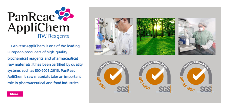
L-Cystine, also known as Diaminoalanine, is a sulfur-containing amino acid that exists in small amounts in proteins and is mostly found in keratin of hair, nails, etc. The structural formula of cystine is as follows:L-Cystine dihydrochloride is the stable salt form of L-cystine, and its structural formula is as follows:L-cystine itself has a very low solubility in water, but its dihydrochloride form greatly enhances water solubility, making it more convenient to use in liquid culture media and drug preparations. Therefore, the function of "L-cystine" discussed is usually realized in practical operation in the form of "L-cystine dihydrochloride". Their applications in biopharmaceuticals mainly fall into two major fields: as key additives in cell culture media and as excipients or stabilizers in drug formulations. 1. As key additive in cell culture mediaThis is the most important and core application of L-cysteine in biopharmaceuticals. ① Provide essential amino acids to support cell growthCells take L-cystine into the cell through transport proteins on their cell membranes, and then rapidly reduce L-cystine to two molecules of L-cysteine in a reducing environment. ② It regulates the REDOX balance and is a precursor for glutathione synthesisL-cysteine is the rate-limiting raw material for the synthesis of glutathione (GSH), the most important antioxidant in cells. GSH can effectively eliminate ROS, protect cells from damage, reduce apoptosis, thereby extending the culture time and increasing the yield of target proteins. ③ Promote the formation of correct disulfide bondsTherapeutic proteins, especially antibodies, rely on correct intramolecular and intermolecular disulfide bond connections for their complex structures. L-cysteine/cysteine, as a direct participant in disulfide bonds, can ensure that disulfide bonds are formed rapidly and accurately during protein synthesis and folding. 2. As an active ingredient in drug preparationsIn addition to being used as an additive in cell culture, L-cystine itself can also be used as a drug or a drug excipient. ① As a therapeutic drugHair and skin health: Cystine is an important component of keratin, which is the main structural protein of hair, skin and nails. Therefore, it is sometimes used as an ingredient in compound preparations to treat hair loss or improve skin conditions.Antioxidant therapy: As a precursor of glutathione, it can theoretically be used to enhance the body's antioxidant capacity, but it is usually used in combination with other drugs (such as B vitamins). ② As an excipient or stabilizer for drug preparationsAntioxidants: By taking advantage of its reducing property, L-cysteine can protect drug components from oxidation and degradation, thereby extending the efficacy period of the drug.Stabilizer: In some specific biopharmaceutical formulations, L-cysteine may help maintain the stability of protein drugs. 3. Advantages of L-Cystine dihydrochlorideStability: Compared with L-cystine, L-cystine dihydrochloride is more stable in both solid state and solution, less prone to moisture absorption and oxidation, and is convenient for storage and weighing.High solubility: It is easy to prepare into a high-concentration stock solution and convenient to add to the culture medium. SummaryL-cystine and L-cystine dihydrochloride are important multifunctional molecules in biopharmaceuticals. Its core value lies in serving as a key additive in cell culture media, by supporting cell growth, maintaining REDOX homeostasis and promoting correct protein folding. At the same time, it also functions as an active ingredient in drugs or an excipient in preparations in a few cases. Meanwhile, the stability and water solubility of L-cysteine dihydrochloride have drawn attention, making it a reliable and indispensable component in biopharmaceutical processes. As a long-term and fixed supplier for many well-known global medium manufacturers and pharmaceutical enterprises, PanReac AppliChem, with its outstanding product quality and strict quality control, provides reliable low-endotoxin pharmaceutical-grade L-cystine and L-cystine dihydrochloride for researchers and pharmaceutical enterprises. CODENAMECAS637765L-Cystine Dihydrochloride low endotoxin, pharma grade30925-07-6Z43645L-Cystine (Ph. Eur.) low endotoxin pure, pharma grade56-89-3 AppliChem Low endotoxin L-cysteine dihydrochloric acid can provide free samples. Welcome to click here to fill in the information and apply for testing. The quantity is limited and it's first come, first served! XMJ is the general agent of PanReac AppliChem in China, providing users with comprehensive technical support and after-sales service. If you are interested in the product, please call Ximeijie customer service hotline 400-050-4006 or visit the website www.gq44.cn for more information. more>Advancing purification strategies for large viral modalities using nanofiber adsorbent technology
Introduction Oncolytic viruses, like herpes simplex virus (HSV) and vaccinia virus (VACV), are gaining traction in cancer therapy due to their selective replication in tumor cells, capacity to induce cell lysis, and ability to trigger anti-tumor immune responses. Similarly, pseudotyped lentiviral vectors (LVV), such as GalV and VSV-G, enhance gene therapy by enabling cell-type-specific transduction. This can can impact downstream purification strategies. Traditional resin-based chromatography approaches face limitations in scalability, yield, and host cell protein (HCP) clearance. To address these bottlenecks, this study investigates a novel nanofiber-based adsorbent as an alternative purification strategy. This innovative platform offers promising advantages in binding capacity, impurity clearance, and process throughput, potentially transforming the downstream processing of large viral vectors. Larger viral vectors for gene therapy: LVV, HSV, and VACV Aims of study ? Evaluate purification performance of a novel nanofiber adsorbent for LVV with different pseudotyped envelopes (VSV-G, 4070A, GalV) and oncolytic virus such as HSV ? Assess host cell driven proteins and dsDNA removal to determine impurity clearance capability ? Investigate scalability potential for industrial implementation of nanofiber-based purification 1. Pseudotyped LVV production and purification workflow 2. Higher binding capacity of VSV-G LVV particles using LentiHERO® nanofiber adsorbent LentiHERO® processed large feed volumes before breakthrough of functional and physical LVV particles. 3. Maximizing the LVV recovery yield with high purity via optimized protocol 82% lentiviral recovery with yields > 2E+9 TU/mL of adsorbent and > 90% dsDNA/HCP clearance 4. Processing with LentiHERO® greatly reduces volumes for formulation at manufacturing scale ? Arginine elution eliminates the need for post-elution dilution, simplifying the process ? Low salt or arginine elution minimizes buffer consumption and reduces processing volume 5. Non-VSV-G pseudotyped LVV purification by LentiHERO®1 Lentiviruses can be pseudotyped with different envelope glycoproteins to enhance transduction efficiency and specific tropism. This impacts downstream purification strategies. ? Efficient purification of non-VSV-G lentiviral vectors (4070A: 100%, GalV: 36%) achieved ? Robust impurity reduction: >98% host cell proteins and >87% residual dsDNA without compromising vector functionality 6. Upstream-downstream workflow of oncolytic HSV-1 7. HSV purification profile on LentiHERO® using wt HSV-1(KOS) Load volume 15 mL to a screening device measured 1.5E+10 IU/mL adsorbent. Infectious HSV recovery yield was 56%, 9.2E+9 IU/mL adsorbent. Most infectious HSV eluted at 0.6 M NaCl. 8. HSV purification profile on LentiHERO® using pBACYAC-HSV-1(KOS) Load volume 20 mL to a screening device measured on average 3E+8 IU/mL adsorbent. Infectious HSV recovery achieved 69%, ~1.4E+8 IU/mL adsorbent by 0.8 M arginine elution with high impurity clearance. Conclusions ? LentiHERO® weak AEX nanofiber effectively purifies a range of viral vectors, including: - Pseudotyped lentiviruses (4070A, VSV-G, GalV) - Oncolytic virus HSV-1 ? High recovery yields achieved: - 4070A: 100%, VSV-G: 82%, GalV: 36%, HSV-1: 69% ? Superior host cell proteins clearance: >97% host cell protein removal ? Scalable and versatile platform suitable for industrial applications ? Potential expansion to other viral vectors like Ad5, Vaccinia, and VLPs ? Supports processing intensification for gene therapy and oncolytic virus manufacturing Product Information Product No. Prodcut Name Package NL100100Nereus LentiHERO2 unit packLH0001001ALentiHERO® 11 mLLH0010001BLentiHERO® 1010 mLLH0010001CLentiHERO® 100100 mL If you are interested in the above products, please click here to fill in the information. Astrea Bioseparations technical experts will provide you with personalized optimization solutions and effectively help you solve the difficult problems in the purification process. Astrea Bioseparations was spun off from the University of Cambridge in 1987 and has over 30 years of experience in the research and development and production of chromatography media. It is a world-class supplier of chromatography media products and services. Currently, more than 21 production processes using Astrea's products have been approved by the FDA and EMA. Astrea Bioseparations has three R&D and production bases worldwide, focusing on providing industry-leading chromatography media and technical services for the fields of biologics and CGT. The new chromatography technology based on nanofibers launched by Astrea Bioseparations has solved the problems of low loading capacity and time-consuming processes of traditional biological separation tools, achieving a faster, more environmentally friendly and cost-effective purification process, fully meeting the needs of today's biopharmaceutical innovation. XMJ Scientific is the agent of Astrea Bioseparations in China, providing users with comprehensive technical support and after-sales service. Welcome to call XMJ's customer service hotline at 400-050-4006 or visit the website www.gq44.cn for more information at any time. more>New Product Launch! Cygnus Recombinant Trypsin Residue Detection Kit
Trypsin is a serine protease that specifically recognizes and cleaves the arginine-lysine (Arg-Lys) peptide bonds in adhesion proteins on the cell surface. It plays a critical role in cell-based vaccine production and gene therapy cell culture, and is widely used to dissociate adherent cells such as HEK293 and Vero, enabling their efficient separation from the surface of culture vessels. However, residual recombinant trypsin in the final product may not only affect process efficiency and product integrity, but also pose potential safety risks. In response to this, the Chinese Pharmacopoeia (2025) has established clear regulatory requirements: · In the general chapter of biological products, Quality Control of Raw Materials and Excipients for Biological Product Production, protein hydrolases of non-animal origin are classified as medium-risk raw materials; · Meanwhile, the General Principles for Human Gene Therapy Products stipulates that potential process-related impurities shall be identified, evaluated, and subject to qualitative and/or quantitative analysis. To accurately monitor and control this critical residual impurity, Cygnus Technologies has launched a new rTrypsin ELISA Detection Kit. Based on the sandwich ELISA principle, this kit features simple operation. During its development, antibodies were prepared using recombinant porcine trypsin and subjected to strict antigen-affinity purification—ensuring high specificity of the antibodies for the target molecule. As such, this kit serves as an ideal process development tool: it can be used to optimize the removal efficiency of recombinant trypsin, as well as for routine quality control and product release testing, helping products meet regulatory requirements.rTrypsin ELISA Kit (Item No.: F1080) Product Performance · Ultra-High Sensitivity: Limit of Detection (LOD) ~0.05 ng/mL; Limit of Quantitation (LOQ) ~0.31 ng/mL · Excellent Precision: Intra-assay and Inter-assay CVs (Coefficients of Variation) are both Cygnus Technologies, LLC. provides products and analytical methods for the biotechnology and biopharmaceutical industries, with the goal of accelerating the R&D phase and enhancing product quality. The company develops and manufactures bioprocess residue detection kits, which are used to detect specific impurities across more than 50 different expression systems. As a specialist in high-sensitivity analytical technologies focused on immunoassays for biotech applications, Cygnus has had its products and services adopted by nearly all major biopharmaceutical companies for over 25 years. As the exclusive distributor of Cygnus in China, Beijing XMJ Technology Co., Ltd. has established long-term and stable cooperative relationships with numerous well-known domestic pharmaceutical companies and CRO/CMO enterprises. Over the years, XMJ’s products and services have helped many enterprises:· Accelerate the R&D phase · Enhance drug quality, purity, and safety · Speed up the optimization of R&D processes · Shorten time-to-market for products · Reduce QC (Quality Control) costs If you are interested in the aforementioned products, please feel free to: · Call XMJ’s customer service hotline: 400-050-4006 · Visit the official website: www.gq44.cn for more information. more>SeraCare?KPL ELISA Substrates with High Cost-Effectiveness
LGC·SeraCare·KPL (hereinafter referred to as KPL), distributed by XMJ, is a world-renowned manufacturer of commercially available affinity-purified secondary antibodies and substrate chromogenic systems. With over 40 years of product R&D experience, KPL has obtained ISO 13485 quality management system certification. Its products feature consistent batch-to-batch performance and reliable quality, earning wide recognition and praise from diagnostic companies worldwide. KPL’s product portfolio includes cost-effective secondary antibodies, substrate chromogenic reagents, and immunoassay auxiliary reagents, which are widely used in immunological research, diagnostic reagent development, and clinical testing. This article will focus on KPL’s high-cost-effectiveness ELISA substrate product series. In ELISA detection, 3,3',5,5'-tetramethylbenzidine (TMB) has become the preferred substrate for horseradish peroxidase (HRP) chromogenic detection, thanks to its high sensitivity, strong absorbance, ease of operation, safety, and broad compatibility. SureBlue? TMB, developed by KPL, is an optimized version of conventional TMB substrates. With over 30 years of application history, it offers higher sensitivity, consistent quality, low batch-to-batch variation, and non-carcinogenic properties. SureBlue Reserve? TMB is a further optimized iteration of SureBlue? TMB. Its detection sensitivity is 50% higher than that of SureBlue? TMB. As a water-based substrate (free of DMF or DMSO), it adopts a ready-to-use single-component formulation for convenient operation and has a shelf life of up to 24 months. Product name Item No. Specification Reaction enzyme Sensitivity SureBlue Reserve? TMB 1-Component Microwell Peroxidase Substrate 5120-00815120-00825120-0083 100 mL 400 mL 1000 mL HRP 5*10-16 SureBlue? TMB 1-Component Microwell Peroxidase Substrate 5120-0075 5120-00765120-00775120-0078 100 mL 100 mL 1000 mL 5000 mL HRP 10-15 TMB 2-ComponentMicrowell Peroxidase Substrate Kit 5120-00535120-00475120-0050 200 mL 600 mL 2700 mL HRP 5*10-15 ABTS® is a universal substrate containing 2,2'-azino-bis(3-ethylbenzothiazoline-6-sulfonic acid). It offers a broad working range, making it suitable for scenarios requiring low background and moderate sensitivity.In the presence of horseradish peroxidase (HRP)-labeled conjugates, ABTS develops a blue-green color at 405–410 nm, and the color remains stable after the reaction is stopped. It is recommended for ELISA applications and is available in both single-component and two-component formats. Product name Item No. Specification Reaction enzyme Sensitivity ABTS® 1-Component Microwell Peroxidase Substrate Kit 5120-00465120-00415120-0043 100 mL 6x100 mL 1000 mL HRP 10-13 ABTS® 2-Component Microwell Peroxidase Substrate Kit 5120-00325120-0033 6x10 mL 6x450 mL HRP 10-13 BluePhos® is a substrate specifically designed for alkaline phosphatase (AP)-labeled conjugates and serves as a high-performance alternative to disodium 4-nitrophenyl phosphate (pNPP). As a soluble, patented form of BCIP, it offers higher sensitivity, a broader working range, lower background signals, and greater ease of use. Supplied in a two-component format, this substrate is ready to use once mixed. It produces a blue product when reacted in microplates or test tubes, making it suitable for both qualitative and quantitative immunoassays. Compared to orange or yellow substrates, it provides clearer visual differentiation. Product name Item No. Specification Reaction enzyme Sensitivity BluePhos® Microwell Substrate(2-C) 5120-0061 5120-0059 5120-0060 50 mL 600 mL 2700 mL AP 10-13 LumiGLO® and LumiGLO Reserve? chemiluminescent substrates are luminol-based substrates specifically designed for use with horseradish peroxidase (HRP)-labeled reporters. LumiGLO Reserve? features enhanced signal intensity, with a sensitivity that is over 20 times higher than that of KPL LumiGLO® and other competing products. Both LumiGLO® and LumiGLO Reserve? chemiluminescent substrates are specialized for detecting low-abundance proteins or proteins derived from precious samples. Product name Item No. Specification Reaction enzyme Sensitivity LumiGLO Reserve? Chemiluminescent Substrate(3-C) 5430-00505430-00515430-0049 600 cm2 1000 cm2 2400 cm2 HRP 10-15-10-12 LumiGLO® Chemiluminescent Substrate(2-C) 5430-00425430-00405430-0041 60 mL 240 mL 720 mL HRP 10-12 Both PhosphaGLO? AP and PhosphaGLO Reserve? AP substrates are designed based on the dioxetane chemiluminescence principle, specifically for use with alkaline phosphatase (AP)-labeled reporters.Among them, PhosphaGLO Reserve? AP substrate delivers a significant boost in signal intensity, making it particularly suitable for detecting low-abundance proteins and target proteins in precious samples.The PhosphaGLO? and PhosphaGLO Reserve? AP substrate series are supplied as stable, single-component solutions. This format simplifies experimental operation while ensuring the stability and reliability of experimental results. Experimental results can be recorded using X-ray film or a chemiluminescence imager to obtain permanent records. For ELISA experiments, it is recommended to use a chemiluminometer for detection to achieve higher sensitivity and accuracy. Product name Item No. Specification Reaction enzyme Sensitivity PhosphaGLO Reserve? AP Substrate(1-C) 5430-00525430-0053 30 mL 100 mL AP 10-15 PhosphaGLO? AP Substrate(1-C) 5430-00545430-0055 30 mL 100 mL AP 10-12 KPL’s substrate product series is highly favored by researchers and diagnostic reagent manufacturers for its exceptional performance. These substrates boast the following key features: · High sensitivity · Consistent batch-to-batch performance · Long shelf life · Flexible format options (single-component or multi-component) · Easy operation · Safety, environmental friendliness, and non-carcinogenicity · Multiple specifications to meet diverse needs In addition to substrates, KPL also offers a full range of auxiliary reagents for experiments including enzyme-linked immunosorbent assay (ELISA), Western blotting (WB), immunohistochemistry (IHC), and enzyme-linked immunospot (ELISpot). These include coating buffers, blocking buffers, washing buffers, and stop solutions. For more information, please feel free to: · Call XMJ’s customer service hotline: 400-050-4006 · Visit the official website: www.gq44.cn With its diverse product options and outstanding performance, KPL’s substrate series has become the preferred brand in the fields of immunological research and diagnostic reagent development. KPL (Kinetic Plasmonics Laboratories) is a company boasting a profound history and an excellent reputation in the biosciences field. As one of the world’s early biotech firms to commercialize affinity-purified secondary antibodies, KPL has laid a solid foundation for its development by virtue of its pioneering status in this domain. Furthermore, KPL is also among the world’s major manufacturers of secondary antibodies and substrate chromogenic systems. With nearly 40 years of experience in product R&D, it has accumulated extensive professional knowledge and strong technical capabilities. The company has obtained ISO 13485 quality management system certification, which ensures minimal batch-to-batch variation and reliable quality for all its products. Characterized by high purity, high sensitivity, and a high signal-to-noise ratio (strong signal, low background), KPL’s products have won the trust of researchers and diagnostic enterprises worldwide. In 2013, KPL was acquired by SeraCare, a supplier of in vitro diagnostic (IVD) reagents, further consolidating its leading position in the industry. Founded in 1984, SeraCare is a key partner for global IVD manufacturers and clinical laboratories, and it became part of the LGC Group in 2018.As the exclusive distributor of the LGC·SeraCare·KPL brand in China, Beijing XMJ is committed to promoting KPL’s high-quality products in the Chinese market and providing premium products and professional services to a large number of domestic researchers. more>Pfanstiehl Secures Its 18th CDE Registration Number for Glycine (G-140)
Warm Congratulations to Pfanstiehl: Injectable-Grade Glycine (G-140) Secures CDE Registration Number F20250000403. This marks the 18th CDE registration number obtained by Pfanstiehl’s products!Glycine is widely used in pharmaceutical excipient formulations, serving as a buffering agent, solubilizer, stabilizer, and more, with numerous advantages. Adhering to its longstanding commitment to high quality, Pfanstiehl has launched its injectable-grade Glycine (G-140) to address the global biopharmaceutical industry’s demand for premium glycine. This product boasts ultra-high purity, ultra-low endotoxins, and ultra-low metal residues, with individual testing conducted for 29 types of metal ions. Furthermore, it incorporates additional quality testing indices, including DNA, RNA, DNase, RNase, lipase, and nitrite. Compliant with the standards of multiple pharmacopoeias (USP, EP, BP, JPE, ChP), it is well-suited for the manufacturing of biotherapeutic drugs and vaccines.Product Information · Product Name: Glycine, High Purity, Low Endotoxin, Low Metals, USP, EP, BP, JPE, ChP (G-140)· Molecular Formula: C?H?NO?· Molecular Weight: 75.07 g/mol· CAS Number: 56-40-6· Item No.: G-140Product Features · Ultra-high purity, ultra-low endotoxins, ultra-low microbial content, and ultra-low residual metal impurities· Additional quality indices for testing: DNA, RNA, DNase, RNase, lipase, and nitrite· Compliance with multiple pharmacopoeias: USP (United States Pharmacopeia), EP (European Pharmacopoeia), BP (British Pharmacopoeia), JPE (Japanese Pharmacopoeia), ChP (Chinese Pharmacopoeia)· Compliance with ICH Q3D guidelines· Compliance with GMP (Good Manufacturing Practice) standards· Obtained DMF (Drug Master File) and CDE (Center for Drug Evaluation) registration numbersProduct Applications · Buffering agent· Stabilizer· SolubilizerIf you are interested in Pfanstiehl’s injectable-grade Glycine (G-140), please contact Beijing XMJ Technology Co., Ltd., Pfanstiehl’s distributor in China, to obtain the following detailed product overview documents and test samples. The latest product series from Pfanstiehl Inc. is as follows. Please feel free to contact Beijing XMJ Technology Co., Ltd. to obtain free 100g samples.Beijing XMJ Technology Co., Ltd. is the authorized distributor of Pfanstiehl in China, providing customers with comprehensive technical support and after-sales service. For more information, please feel free to call XMJ’s customer service hotline at 400-050-4006 or visit the official website at www.gq44.cn. more>New Product Launch | Alzet Osmotic Pump 100μL Sustained Release for 6 Weeks (Model 1006)
As the latest addition to its osmotic pump product line, the new Model 1006 from Alzet enables continuous drug delivery for up to 6 weeks, making it the osmotic pump with the longest sustained release duration among Alzet pumps of the 100μL . Model 1006 sets a new industry standard for small-dose, long-term animal drug delivery applications.Designed specifically for preclinical research, this product delivers precise and stable infusion of various formulations, including drugs, hormones, and other compounds. It serves as an ideal solution for chronic research projects that require long-term maintenance of a constant infusion rate.Key Advantages of the Alzet Model 1006 Pump:· 6-Week Sustained Drug Delivery: Currently the longest sustained release duration among Alzet pumps of the 100μL size.· Compact Size: Suitable for mice and other small animal models.· Precise Infusion: Ensures reliable and stable sustained release rates.Specifications of the Alzet Model 1006 Pump:Delivery Volume:100μlSustained Release Rate:0.08μl/hContinuous Delivery Duration:6 WeeksPriming Time:72hAlzet Osmotic Pump Product ListModelProduct NameSpecification1003DImplantable Capsule Osmotic Pump, 100μL, 3-Days Sustained Release10 pcs/box1007DImplantable Capsule Osmotic Pump, 100μL, 7-Days Sustained Release10 pcs/box1002Implantable Capsule Osmotic Pump, 100μL, 2-weeks Sustained Release10 pcs/box1004Implantable Capsule Osmotic Pump, 100μL, 4-weeks Sustained Release10 pcs/box1006Implantable Capsule Osmotic Pump, 100μL, 2-weeks Sustained ReleaseNEW10 pcs/box2001DImplantable Capsule Osmotic Pump, 200μL, 1-Day Sustained Release10 pcs/box2001Implantable Capsule Osmotic Pump, 200μL, 1-Week Sustained Release10 pcs/box2002Implantable Capsule Osmotic Pump, 200μL, 2-Weeks Sustained Release10 pcs/box2004Implantable Capsule Osmotic Pump, 200μL, 4-Weeks Sustained Release10 pcs/box2006Implantable Capsule Osmotic Pump, 200μL, 6-Weeks Sustained Release10 pcs/box2ML1Implantable Capsule Osmotic Pump, 2000μL, 1-Week Sustained Release10 pcs/box2ML2Implantable Capsule Osmotic Pump, 2000μL, 2-Week2 Sustained Release10 pcs/box2ML4Implantable Capsule Osmotic Pump, 2000μL, 4-Weeks Sustained Release10 pcs/boxBeijing XMJ Technology Co., Ltd. is the authorized Level-1 distributor of Alzet in China, providing you with comprehensive pre-sales and after-sales technical support.If you need more information about Alzet sustained-release osmotic pumps, please feel free to:· Call XMJ’s national customer service hotline: 400-050-4006· Visit XMJ’s official website: www.gq44.cn for inquiries and orders more>Guide to HCP ELISA Plate Layout Optimization: Key Steps to Improve Data Accuracy
To date, the ELISA method remains the gold standard for detecting and quantifying Host Cell Proteins (HCPs) in process monitoring and product batch release. Cygnus has been providing HCP detection kits for over 25 years, and has developed numerous patented methods to produce antibodies with broad reactivity against HCPs. Currently, it offers more than 32 types of HCP ELISA detection kits tailored to different expression platforms. Cygnus' ELISA kits utilize pre-coated plates, where antigens and detection antibodies are added simultaneously for incubation during the assay. In reality, HCP detection is quite complex, as it primarily relies on HCP antibodies capable of reacting with thousands of HCP species. For ELISA results to be accurate, both the capture antibodies and detection antibodies targeting the antigens must be in excess. Only under the condition of excess antibodies can a dose-response curve with a positive slope be obtained, thereby enabling accurate quantification. Therefore, a rational plate layout setup is an excellent starting point. Basic Procedure for Cygnus HCP ELISA Assay I. HCP ELISA Plate Layout Specifications 1. Requirement for Technical Replicates A triplicate well design is recommended, as it is the most effective approach. It allows for the identification and elimination of potential outliers and provides more information when troubleshooting issues after the experiment. It should be noted that if the number of test samples does not support triplicate wells, at least duplicate wells must be set up—detection without any technical replicates is considered invalid. If possible, it is preferable to set up triplicate wells for both standards and controls. 2. Blank Control Setting up additional blank controls is not recommended. Subtracting blank control values will not improve experimental results; in fact, it may even lead to an increase in the Coefficient of Variation (CV). For details, please refer to the document How Should Blank Controls Be Set Up When Using Cygnus ELISA Kits?. 3. Setting Up Positive Controls Use a standard within the mid-concentration range of the standard curve as the positive control (low-concentration standards are more affected by assay sensitivity). When using a positive control derived from HCP standards, the mid-concentration point of the curve typically provides the most information. 4. Setting Up Negative Controls Use sample dilution buffer as the negative control to evaluate whether there is a significant deviation between the 0-concentration standard and the dilution buffer used. 5. Setting Up 1–2 Additional HCP Controls A well-characterized in-process sample from the manufacturing workflow can be used as an additional HCP control. If there are extra wells available, setting up two dilutions of this sample is more effective than using only one dilution. 6. Optimizing Sample Loading with Low-Bind Plates When loading samples into the well, the most important considerations are: how long each step will take and the direction of movement for each step. To address this, pre-aliquot the test samples and enzyme-conjugated secondary antibodies into a low-bind plate first, then transfer the entire volume to the assay plate. This method significantly reduces sample loading errors when adding samples to the assay plate. II. Example of ELISA Plate Layout The optimal way to arrange samples is to place standards on the left side of the plate and controls (negative or positive) on the right side. Meanwhile, factors such as the number of technical replicates required, sample dilution gradients, and the direction of movement during sample loading and plate washing must also be considered. By placing standards and quality control samples on opposite sides of the plate, you ensure that the same information can be obtained from either side of the plate. This clearly reveals any operational errors that may occur during sample loading or plate washing, and adjusting the positions of these wells within the plate enables more effective troubleshooting. 1. Recommended Schematic for Triplicate Well Layout For 5 samples with 4 dilution gradients: all standards, samples, and controls are set up in triplicate wells.2. Recommended Schematic for Duplicate Well Plate Layout For 4 samples and spiked control samples, 4 dilution gradients are set for each. All standards, samples, and control samples are arranged in duplicate wells. 3. Recommended Schematic for Plate Layout of HCP Samples with Unknown Concentrations For 4 samples, 8 dilution gradients are set up to collect most of the information about samples with unknown HCP levels. Additional plate layout examples can be found at the end of the document. III. Troubleshooting and Solutions for Common HCP ELISA Plate Layout Issues 1. Sample Loading Problems During the HCP ELISA assay, every 2 minutes of static incubation is equivalent to 1 minute of shaking incubation. In other words, if sample loading takes 20 minutes, the samples added first will have an incubation time 10 minutes longer than those added last. Suppose the shaking incubation time is set to 1 hour, while sample loading alone takes 20 minutes—this will inevitably lead to differences in binding efficiency between the samples added earlier and later. Specifically, the samples added first will show abnormally high Optical Density (OD) values. Therefore, the core principle of sample loading is to ensure that the incubation time is as consistent as possible across all wells in the plate. If a large number of samples need to be tested at one time, to avoid excessive time spent on sample loading, we recommend pre-adding the samples and enzyme-conjugated antibodies into a low-bind plate first, then quickly transferring them to the assay plate using a multi-channel pipette. Practice has proven that this operation method can effectively resolve result abnormalities caused by differences in sample loading time. 2. Plate Washing Problems Improper plate washing operations can also lead to abnormalities in assay results. Similar to sample loading, the core principle to follow for HCP ELISA plate washing is alternating-direction washing—that is, ensuring the total washing time is as consistent as possible across all wells. For precautions regarding plate washing operations, please refer to the document How to Resolve ELISA Assay Result Drift with Correct Plate Washing Methods?. IV. Common Error Cases and Improvement Methods Case 1: Errors Caused by Slow Sample Loading If the same sample is loaded slowly into each well in the direction from Row A to Row H and Column 1 to Column 12 sequentially, this loading method will affect the assay results—even if the correct plate washing method is adopted. The following heatmap example shows what happens when the same sample is added to the microplate at a slow speed. 1 2 3 4 5 6 7 8 9 10 11 12 A 1.000 0.976 0.952 0.928 0.904 0.880 0.856 0.832 0.808 0.784 0.760 0.736 B 0.997 0.973 0.949 0.925 0.901 0.877 0.853 0.829 0.805 0.781 0.757 0.733 C 0.994 0.970 0.946 0.922 0.898 0.874 0.850 0.826 0.802 0.778 0.754 0.730 D 0.991 0.967 0.943 0.919 0.895 0.871 0.847 0.823 0.799 0.775 0.751 0.727 E 0.988 0.964 0.940 0.916 0.892 0.868 0.844 0.820 0.796 0.772 0.748 0.724 F 0.985 0.961 0.937 0.913 0.889 0.865 0.841 0.817 0.793 0.769 0.745 0.721 G 0.982 0.958 0.934 0.910 0.886 0.862 0.838 0.814 0.790 0.766 0.742 0.718 H 0.979 0.955 0.931 0.907 0.883 0.859 0.835 0.811 0.787 0.763 0.739 0.715 Improvement Method: First, pre-add the enzyme-conjugated antibodies and samples into a low-bind plate. Then, use a multi-channel pipette to quickly transfer all samples to the assay plate for incubation. This method can effectively reduce the time difference in sample loading between different wells, ensure the incubation time of all wells is as consistent as possible, and thereby reduce the probability of outliers. Case 2: Errors Caused by Plate Washing in the Same Direction If the washing buffer is added in the same direction for all 4 washing cycles, it will also affect the assay results. The following heatmap example shows what may happen when the washing buffer is added in the direction from Column 1 to Column 12 using a multi-channel pipette for all 4 plate washing cycles. 1 2 3 4 5 6 7 8 9 10 11 12 A 0.670 0.700 0.730 0.760 0.790 0.820 0.850 0.880 0.910 0.940 0.970 1.000 B 0.670 0.700 0.730 0.760 0.790 0.820 0.850 0.880 0.910 0.940 0.970 1.000 C 0.670 0.700 0.730 0.760 0.790 0.820 0.850 0.880 0.910 0.940 0.970 1.000 D 0.670 0.700 0.730 0.760 0.790 0.820 0.850 0.880 0.910 0.940 0.970 1.000 E 0.670 0.700 0.730 0.760 0.790 0.820 0.850 0.880 0.910 0.940 0.970 1.000 F 0.670 0.700 0.730 0.760 0.790 0.820 0.850 0.880 0.910 0.940 0.970 1.000 G 0.670 0.700 0.730 0.760 0.790 0.820 0.850 0.880 0.910 0.940 0.970 1.000 H 0.670 0.700 0.730 0.760 0.790 0.820 0.850 0.880 0.910 0.940 0.970 1.000 Improvement Method:It is recommended to perform plate washing in alternating directions—specifically, add the washing buffer from opposite directions for the 1st and 3rd washing cycles versus the 2nd and 4th washing cycles. V. Cases of Abnormal HCP ELISA Results and TroubleshootingAs shown in the figure below, the test results exhibit a decreasing trend from left to right among the technical replicates of the samples. Additionally, the Optical Density (OD) values of both the negative controls and positive controls on the right side of the plate are lower than those on the left side. The inconsistency between the positive control results on the two sides indicates that this experiment is a failure. Furthermore, the average difference in OD values among the negative control replicates on the left side is 0.01, while the OD values of the negative controls on the right side are close to the background level. Therefore, additional information is required to further determine whether the issue stems from sample loading or plate washing. In summary, the possible causes of the failure in this experiment are: 1. Slow sample loading;2. Failure to perform plate washing in alternating directions. 1 2 3 4 5 6 7 8 9 10 11 12 A STD 1 Sample 1 Dilution 1 Sample 3 Dilution 1 Sample 5 Dilution 1 B STD 2 Sample 1 Dilution 2 Sample 3 Dilution 2 Sample 5 Dilution 2 C STD 3 Sample 1 Dilution 3 Sample 3 Dilution 3 Sample 5 Dilution 3 D STD 4 Sample 1 Dilution 4 Sample 3 Dilution 4 Sample 5 Dilution 4 E STD 5 Sample 2 Dilution 1 Sample 4 Dilution 1 Negative Control (Diluent) F STD 6 Sample 2 Dilution 2 Sample 4 Dilution 2 Control Dilution 1(External HCP) G STD 7 Sample 2 Dilution 3 Sample 4 Dilution 3 Control Dilution 2 (External HCP) H STD 8 Sample 2 Dilution 4 Sample 4 Dilution 4 Postive Control (STD 5 repeat) 1 2 3 4 5 6 7 8 9 10 11 12 A 2.500 2.450 2.400 1.200 1.150 1.100 2.000 1.950 1.900 1.700 1.650 1.600 B 1.200 1.150 1.100 0.600 0.550 0.500 1.000 0.950 0.900 0.850 0.800 0.750 C 0.600 0.550 0.500 0.300 0.280 0.260 0.500 0.480 0.460 0.425 0.405 0.385 D 0.300 0.250 0.200 0.150 0.140 0.130 0.250 0.240 0.230 0.212 0.202 0.192 E 0.150 0.130 0.110 1.400 1.350 1.300 0.125 0.115 0.105 0.005 0.005 0.005 F 0.075 0.065 0.055 0.700 0.650 0.600 0.550 0.500 0.450 0.230 0.225 0.220 G 0.038 0.033 0.028 0.350 0.330 0.310 0.290 0.270 0.250 0.115 0.095 0.075 H 0.015 0.010 0.005 0.175 0.165 0.155 0.145 0.135 0.125 0.090 0.080 0.070 Summaryl A rational microplate layout should be planned before conducting an ELISA assay: an optimized microplate layout enables the monitoring of key performance indicators and faster troubleshooting when issues arise. For more plate layout examples, please click here to access them.l Key considerations for HCP ELISA include: setup of technical replicates, sample dilution gradients, sample loading and plate washing operations, and setup of controls.l Optimizing sample loading operations with low-bind plates can minimize deviations in Optical Density (OD) values.l Do not use blank controls, as this practice will increase the coefficient of variation (CV) of the assay results. Related product recommendations:Product Name:Sample Treatment Plate (Sample processing plate)Item No.:F402Specification:1 plate Cygnus Technologies, LLC. provides products and analytical methods for the biotechnology and biopharmaceutical industries, aiming to accelerate the research and development (R&D) phase and enhance product quality.Cygnus develops and manufactures bioprocess residual test kits, which are used to detect specific impurities in more than 50 different expression systems.As an expert in high-sensitivity analytical technologies focused on immunoassays for biotechnological applications, Cygnus has had its products and services adopted by nearly all major biopharmaceutical companies for over 25 years. As the exclusive general distributor of Cygnus in China, Beijing XMJ Technology Co., Ltd. has established long-term and stable cooperative relationships with numerous well-known domestic pharmaceutical companies and CRO/CMO enterprises. more>About Us
- Resouces
- Research products
- Microbiology & cell culture
- Purification
- Quality control products
- Excipients
- Diagnostic raw materials
- Instruments, equipment and consumables
- Technical service
- Downloads
- Contact Us
- Headquarters:B-10F Golden Resources Business Center, No.2 Landianchang East Road, Haidian District, Beijing, China
- Phone: +86 10-8859-7838
Toll Free Number: +86 400-050-4006
Email: info@xmjsci.com
.png) 京公網(wǎng)安備 11010802028692號
京公網(wǎng)安備 11010802028692號