BOUTIQUEResouces
The occurrence and development of tumors is a complex process involving a series of genetic and epigenetic changes. These changes enable cells to break free from the constraints of normal growth regulatory signals (such as external environmental and internal signals) and acquire the ability for autonomous proliferation. Among these changes, anchorage-independent growth is one of the important markers of malignant cell transformation, and the Soft Agar Colony Formation Assay is regarded as the gold standard for detecting malignant cell transformation.
The Soft Agar Colony Formation Assay cultures cells in a semi-solid medium and observes whether they can form colonies under anchorage-independent conditions, thereby determining the cells’ malignant transformation ability. However, this method usually takes 3–4 weeks to yield results, which seriously slows down research progress. Additionally, since it relies on manual microscopic counting of colonies, unified judgment is difficult to achieve, leading to result deviations. Most importantly, the traditional soft agar assay cannot recover viable cells, which limits the conduct of subsequent research.
Cellbiolabs’ new-generation CytoSelect? Assay System combines fluorescence quantification technology and an improved soft agar formulation, comprehensively optimizing the experimental workflow:
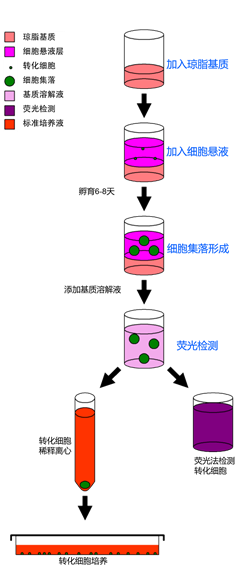
The CytoSelect? Cell Transformation Assay Kit quantifies cell colonies via a high-sensitivity fluorescence detection method. It shortens the experimental cycle from 3–4 weeks to within 1 week, significantly improving research efficiency while avoiding subjective errors caused by manual counting. This kit is particularly suitable for high-throughput sample detection. For application fields such as protein/DNA microarray analysis or cancer vaccine development, the use of a dedicated, improved soft agar medium enables easy recovery of activated transformed cells for further culture and testing.
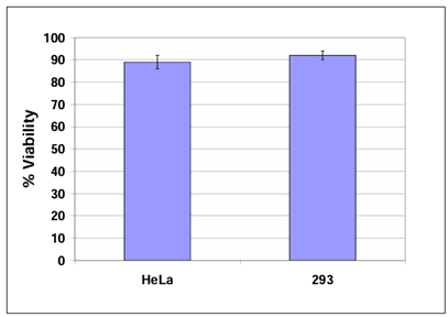
Fig. 1 Cell Viability Assay:HeLa and 293 cells were recovered and cultured for 6 days in accordance with the experimental protocol, and cell viability was determined using the trypan blue exclusion method.
Product Ordering Information:
|
Item Number |
Product Name |
Test Method |
|
CBA-135 |
CytoSelect? 96-Well Cell Transformation Assay, Cell Recovery Compatible |
Colorimetric method |
|
CBA-140 |
CytoSelect? 96-Well Cell Transformation Assay, Cell Recovery Compatible |
Fluorescence method |
|
CBA-130 |
CytoSelect? 96-Well Cell Transformation Assay, Soft Agar Colony Formation |
Fluorescence method |
Published Literature on Product-Related Applications
1. El Baba, R. et al. (2023). Polyploidy, EZH2 upregulation, and transformation in cytomegalovirusinfected human ovarian epithelial cells. Oncogene. doi: 10.1038/s41388-023-02813-4.
2. Hiroki, H. et al. (2023). Targeting Poly(ADP)ribose polymerase in BCR/ABL1-positive cells. SciRep. 13(1):7588. doi: 10.1038/s41598-023-33852-2.
3. El Baba, R. et al. (2023). EZH2-Myc driven glioblastoma elicited by cytomegalovirus infection ofhuman astrocytes. Oncogene. doi: 10.1038/s41388-023-02709-3.
4. Kantisin, S. et al. (2022). In utero arsenic exposure increases DNA damage and gene expressionchanges in umbilical cord mesenchymal stem cells (UC-MSCs) from newborns as well as in UCMSC differentiated hepatocytes. Toxicol Rep. doi: 10.1016/j.toxrep.2022.09.002.
5. Nehme, Z. et al. (2022). Polyploid giant cancer cells, EZH2 and Myc upregulation in mammaryepithelial cells infected with high-risk human cytomegalovirus. EBioMedicine. 80:104056. doi:10.1016/j.ebiom.2022.104056.
6. Kim, D.G. et al. (2022). AIMP2-DX2 provides therapeutic interface to control KRAS-driventumorigenesis. Nat Commun. 13(1):2572. doi: 10.1038/s41467-022-30149-2.
7. Buranarom, A. et al. (2021). Dichloromethane increases mutagenic DNA damage andtransformation ability in cholangiocytes and enhances metastatic potential in cholangiocarcinomacell lines. Chem Biol Interact. doi: 10.1016/j.cbi.2021.109580.
8. Nehme, Z. et al. (2021). Polyploid giant cancer cells, stemness and epithelial-mesenchymalplasticity elicited by human cytomegalovirus. Oncogene. doi: 10.1038/s41388-021-01715-7.
9. Andrade, F. et al. (2021). Polymeric micelles targeted against CD44v6 receptor increaseniclosamide efficacy against colorectal cancer stem cells and reduce circulating tumor cells in vivo.J Control Release. 331:198-212. doi: 10.1016/j.jconrel.2021.01.022.
10. Wakae, K. et al. (2020). EBV-LMP1 induces APOBEC3s and mitochondrial DNA hypermutationin nasopharyngeal cancer. Cancer Med. doi: 10.1002/cam4.3357.
11. Lv, W. et al. (2020). Reprogramming of Ovarian Granulosa Cells by YAP1 Leads to Developmentof High-Grade Cancer with Mesenchymal Lineage and Serous Features. Sci Bull. doi:10.1016/j.scib.2020.03.040.
12. Murata, M. et al. (2020). OVOL2-Mediated ZEB1 Downregulation May Prevent Promotion ofActinic Keratosis to Cutaneous Squamous Cell Carcinoma. J Clin Med. 9(3). pii: E618. doi:10.3390/jcm9030618.
13. Hernandez, D.M. et al. (2020). IPF pathogenesis is dependent upon TGFβ induction of IGF-1.FASEB J. doi: 10.1096/fj.201901719RR.
14. Sand, A. et al. (2019). WEE1 inhibitor, AZD1775, overcomes trastuzumab resistance by targetingcancer stem-like properties in HER2-positive breast cancer. Cancer Lett. 472:119-131. doi:10.1016/j.canlet.2019.12.023.
15. Paul, M. et al. (2022). Nitric-Oxide Synthase trafficking inducer (NOSTRIN) is an emerging negative regulator of colon cancer progression. BMC Cancer. 22(1):594. doi: 10.1186/s12885-022-09670-6.
16. Kondo, M. et al. (2021). Safety and efficacy of human juvenile chondrocyte-derived cell sheets forosteochondral defect treatment. NPJ Regen Med. 6(1):65. doi: 10.1038/s41536-021-00173-9.
17. van der Toorn, M. et al. (2018). The biological effects of long-term exposure of human bronchialepithelial cells to total particulate matter from a candidate modified-risk tobacco product. Toxicol In Vitro. 50:95-108. doi: 10.1016/j.tiv.2018.02.019.
18. Montalbano, M. et al. (2016). Modeling of hepatocytes proliferation isolated from proximal and distal zones from human hepatocellular carcinoma lesion. PLoS One. 11:e0153613.
19. Choi, B.Y. et al. (2023). Engineered Mesenchymal Stem Cells Over-Expressing BDNF Protect theBrain from Traumatic Brain Injury-Induced Neuronal Death, Neurological Deficits, and CognitiveImpairments. Pharmaceuticals (Basel). 16(3):436. doi: 10.3390/ph16030436.
20. Ikeda, J. et al. (2023). Hypoxia inducible factor‐1 activator munc‐18‐interacting protein 3 promotestumour progression in urothelial carcinoma. Clin Transl Disc. 3:e158. doi: 10.1002/ctd2.158.
21. Switzer, C.H. et al. (2022). NOS2 and S-nitrosothiol signaling induces DNA hypomethylation andLINE-1 retrotransposon expression. Proc Natl Acad Sci U S A. 119(21):e2200022119. doi:10.1073/pnas.2200022119.
22. Furuya, K. et al. (2022). Machine learning extracts oncogenic-specific γ-H2AX foci formationpattern upon genotoxic stress. Genes Cells. doi: 10.1111/gtc.13005.
23. Kim, M. et al. (2022). BRAFV600E Mutation Enhances Estrogen-Induced Metastatic Potential ofThyroid Cancer by Regulating the Expression of Estrogen Receptors. Endocrinol Metab (Seoul).37(6):879-890. doi: 10.3803/EnM.2022.1563.
24. Toh, P.J.Y. et al. (2022). Optogenetic control of YAP cellular localisation and function. EMBORep. doi: 10.15252/embr.202154401.
25. Lee, A.R. et al. (2022). Biomarker LEPRE1 induces pelitinib-specific drug responsiveness byregulating ABCG2 expression and tumor transition states in human leukemia and lung cancer. SciRep. 12(1):2928. doi: 10.1038/s41598-022-06621-w.
26. Wang, Y. et al. (2022). Long non-coding RNA OIP5-AS1 suppresses microRNA-92a to augmentproliferation and metastasis of ovarian cancer cells through upregulating ITGA6. J Ovarian Res.15(1):25. doi: 10.1186/s13048-021-00937-3.
27. Andriolo, G. et al. (2021). GMP-Grade Methods for Cardiac Progenitor Cells: Cell BankProduction and Quality Control. Methods Mol Biol. doi: 10.1007/7651_2020_286.
28. Tan, T.T. et al. (2021). Assessment of Tumorigenic Potential in Mesenchymal-Stem/Stromal-CellDerived Small Extracellular Vesicles (MSC-sEV). Pharmaceuticals. 14(4):345. doi:10.3390/ph14040345.
29. Lo, E.K.K. et al. (2021). Low dose of zearalenone elevated colon cancer cell growth through Gprotein-coupled estrogenic receptor. Sci Rep. 11(1):7403. doi: 10.1038/s41598-021-86788-w.
30. Park, S. et al. (2021). Cerebral Cavernous Malformation 1 Determines YAP/TAZ SignalingDependent Metastatic Hallmarks of Prostate Cancer Cells. Cancers (Basel). 13(5):1125. doi:10.3390/cancers13051125.
References
1. El Baba, R. et al. (2023). Polyploidy, EZH2 upregulation, and transformation in cytomegalovirus infected human ovarian epithelial cells. Oncogene. doi: 10.1038/s41388-023-02813-4.
2. Hiroki, H. et al. (2023). Targeting Poly(ADP)ribose polymerase in BCR/ABL1-positive cells. Sci Rep. 13(1):7588. doi: 10.1038/s41598-023-33852-2.
3. El Baba, R. et al. (2023). EZH2-Myc driven glioblastoma elicited by cytomegalovirus infection of human astrocytes. Oncogene. doi: 10.1038/s41388-023-02709-3.
4. Kantisin, S. et al. (2022). In utero arsenic exposure increases DNA damage and gene expression changes in umbilical cord mesenchymal stem cells (UC-MSCs) from newborns as well as in UC-MSC differentiated hepatocytes. Toxicol Rep. doi: 10.1016/j.toxrep.2022.09.002.
5. Nehme, Z. et al. (2022). Polyploid giant cancer cells, EZH2 and Myc upregulation in mammary epithelial cells infected with high-risk human cytomegalovirus. EBioMedicine. 80:104056. doi: 10.1016/j.ebiom.2022.104056.

Cell Biolabs
Headquartered in San Diego, California, USA, Cell Biolabs has been committed to developing technologies and tools for the life sciences research field and commercializing the innovative technological achievements it has developed. The company diligently refines its products with the aim of advancing the research on cell functions and disease mechanisms to new heights.
Cell Biolabs’ products feature unique characteristics and stand at the cutting-edge of the industry. They are widely used in research laboratories across numerous universities, government research institutions, as well as biotechnology and pharmaceutical companies around the world.

Beijing XMJ is the authorized distributor of the Cell Biolabs brand in China, providing users with comprehensive technical support and after-sales service. If you are interested in the products, please feel free to call XMJ's customer service hotline at 400-050-4006 or visit the website www.gq44.cn for more information.
.png) 京公網(wǎng)安備 11010802028692號
京公網(wǎng)安備 11010802028692號